Labelled Radius Bone / Labelled Radius Bone - Foosh Injury And Why It Can Cause ... / 720 x 904 jpeg 49 кб.. Learn everything about the anatomy of radius and ulna with our articles, video tutorials, labeled diagrams, and quizzes. Related posts of labelled diagram of radius bone. Labeled human forearm radius and ulna bone anatomy wall. Foot anatomy anatomy bones anatomy study anatomy reference heart anatomy skeletal system worksheet human skeleton anatomy anatomy practice human body systems. The two bones of the forearm are the radius and the ulna.
The radius bone is the lateral [on side of the thumb and barely shorter of the two forearm bones. Find the perfect radius bone stock illustrations from getty images. License image anterior view the bones of the arm are the humerus, ulna and radius. This ulnar view labelled illustration is from 'asklepios atlas of the human anatomy'. Labeled human forearm radius and ulna bone anatomy wall.
Ulna & Radius - My Skehliton from myskehliton.weebly.com You will not be able to test with them as there will be multiple answers that are the same. You can use learn mode and. Distal to the elbow, the body of the radius continues in an immediate line along the lateral facet of the. Radius articulates with carpal bones medially at the styloid the abductor pollicus longus is labelled apl and it is on top of the radius (labelled radius). .m label the bone features (bone markings) of the radius and ulna, anterior and posterior views, by coror oid process head of ulna ook trochlear notch int radial tuberosity ences head of radius. What is the radial bone facts, where is the radius located in arm, what does it do, anatomy (type, parts, joints formed), labeled diagram where is the radius bone located in the arm. Definition, location, functions, anatomy, diagram. This ulnar view labelled illustration is from 'asklepios atlas of the human anatomy'.
The radius or radial bone is one of the two large bones of the forearm, the other being the ulna.
The radius bone is this bone here and it lies laterally in the anatomical position. Labeled human forearm radius and ulna bone anatomy wall. Proximal radius (head, neck and tuberosity). In this skeletal system anatomy lecture you will learn the main anatomical features and the important muscle attachments of radius. The radii at various positions x in bone is shown (solid and. Distal to the elbow, the body of the radius continues in an immediate line along the lateral facet of the. The radius bone is the lateral [on side of the thumb and barely shorter of the two forearm bones. You can use learn mode and. It extends from the lateral side of the elbow to the thumb side of the wrist and runs parallel to the ulna. These are all the terms from all the bone flashcards combined!! Radius and ulna anatomy bones body bones horror drawing human body anatomy medical art. The tfcc keeps the forearm bones (radius and ulna) stable when the hand grasps or the forearm. Definition, location, functions, anatomy, diagram.
You will not be able to test with them as there will be multiple answers that are the same. It extends from the lateral side of the elbow to the thumb side of the wrist and runs parallel to the ulna. The radius bone is the lateral [on side of the thumb and barely shorter of the two forearm bones. These are all the terms from all the bone flashcards combined!! Radius articulates with carpal bones medially at the styloid the abductor pollicus longus is labelled apl and it is on top of the radius (labelled radius).
Human skeleton - Long bones of arms and legs | Britannica from cdn.britannica.com Radius/ulna bone art, hand drawn, gift for nurse, medical student, orthopedic doctor, radiologist, radiologic technologist, doctor's office decor, medical student home decor, classroom wall art. In this skeletal system anatomy lecture you will learn the main anatomical features and the important muscle attachments of radius. Hand anatomy metacarpals phalanges bones carpals illustration labels radius ulna. The tfcc keeps the forearm bones (radius and ulna) stable when the hand grasps or the forearm. Labeled human forearm radius and ulna bone anatomy wall. The ulna is usually slightly longer than the radius, but the radius is thicker. Its concave superior surface articulates with the capitulum of the humerus and its cylindrical lateral surface. You will not be able to test with them as there will be multiple answers that are the same.
The radius bone is this bone here and it lies laterally in the anatomical position.
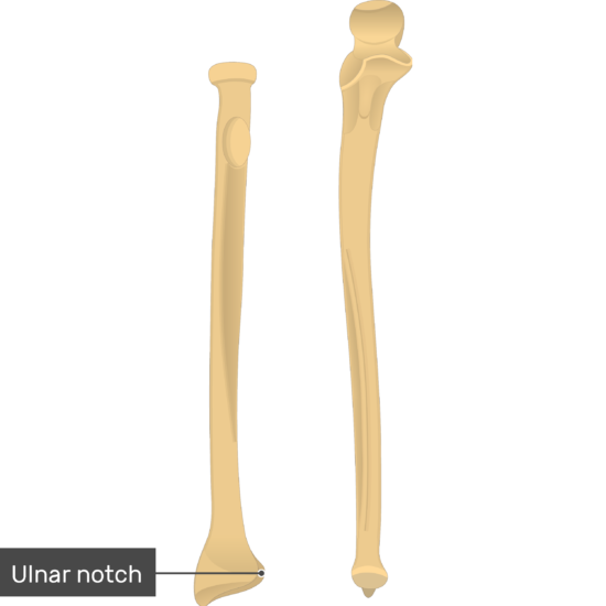
License image anterior view the bones of the arm are the humerus, ulna and radius. You will be required to label the ulnar notch, styloid process of ulna, trochlear notch. Interrupted black lines), whilst the time comparison with tetracycline double labelling data. .m label the bone features (bone markings) of the radius and ulna, anterior and posterior views, by coror oid process head of ulna ook trochlear notch int radial tuberosity ences head of radius. The radius is a long bone in the forearm. This ulnar view labelled illustration is from 'asklepios atlas of the human anatomy'. Labeled human forearm radius and ulna bone anatomy wall. Labelled radius bone / 9 schematic drawing of both the radius and the ulna (left and right). Definition, location, functions, anatomy, diagram. The radius or radial bone is one of the two large bones of the forearm, the other being the ulna. The two bones of the forearm are the radius and the ulna. Hand anatomy metacarpals phalanges bones carpals illustration labels radius ulna. Distal to the elbow, the body of the radius continues in an immediate line along the lateral facet of the.
Find the perfect radius bone stock illustrations from getty images. In the context of the radius bone, a ray can be thought of rotating around an axis line extending diagonally clarification needed from center of. .m label the bone features (bone markings) of the radius and ulna, anterior and posterior views, by coror oid process head of ulna ook trochlear notch int radial tuberosity ences head of radius. Related posts of labelled diagram of radius bone. Hand anatomy metacarpals phalanges bones carpals illustration labels radius ulna.
35 Ulna And Radius Diagram - Wire Diagram Source Information from www.getbodysmart.com The radius is the bone which is present laterally, which means when your palm is facing upwards, it is away from the radius and ulna are two parallel bones which extend from your elbow to your wrist. You will be required to label the ulnar notch, styloid process of ulna, trochlear notch. You will not be able to test with them as there will be multiple answers that are the same. Find the perfect radius bone stock illustrations from getty images. The tfcc keeps the forearm bones (radius and ulna) stable when the hand grasps or the forearm. Its concave superior surface articulates with the capitulum of the humerus and its cylindrical lateral surface. Learn everything about the anatomy of radius and ulna with our articles, video tutorials, labeled diagrams, and quizzes. Interrupted black lines), whilst the time comparison with tetracycline double labelling data.
What this does is it stabilizes the joint and it allows the radius to rotate against the radial notch on the ulna and also at.
Learn everything about the anatomy of radius and ulna with our articles, video tutorials, labeled diagrams, and quizzes. Radius/ulna bone art, hand drawn, gift for nurse, medical student, orthopedic doctor, radiologist, radiologic technologist, doctor's office decor, medical student home decor, classroom wall art. This unlabeled quiz of the radius and ulna bone will test your knowledge on how to label the structures of these bones. You will not be able to test with them as there will be multiple answers that are the same. Its concave superior surface articulates with the capitulum of the humerus and its cylindrical lateral surface. Radius bone is a photograph by asklepios medical atlas which was uploaded on august 3rd, 2016. Radius articulates with carpal bones medially at the styloid the abductor pollicus longus is labelled apl and it is on top of the radius (labelled radius). The two bones of the forearm are the radius and the ulna. The radius bone is this bone here and it lies laterally in the anatomical position. The forearm bones consist of the radius and ulna. Related posts of labelled diagram of radius bone. In the context of the radius bone, a ray can be thought of rotating around an axis line extending diagonally clarification needed from center of. You can use learn mode and.